There are different types of stains, namely gram stain, capsule stain, endospore stain and acid fast stain. Only acid fast stain is described in this procedure. Click on the other types of staining below to learn about them.
Introduction
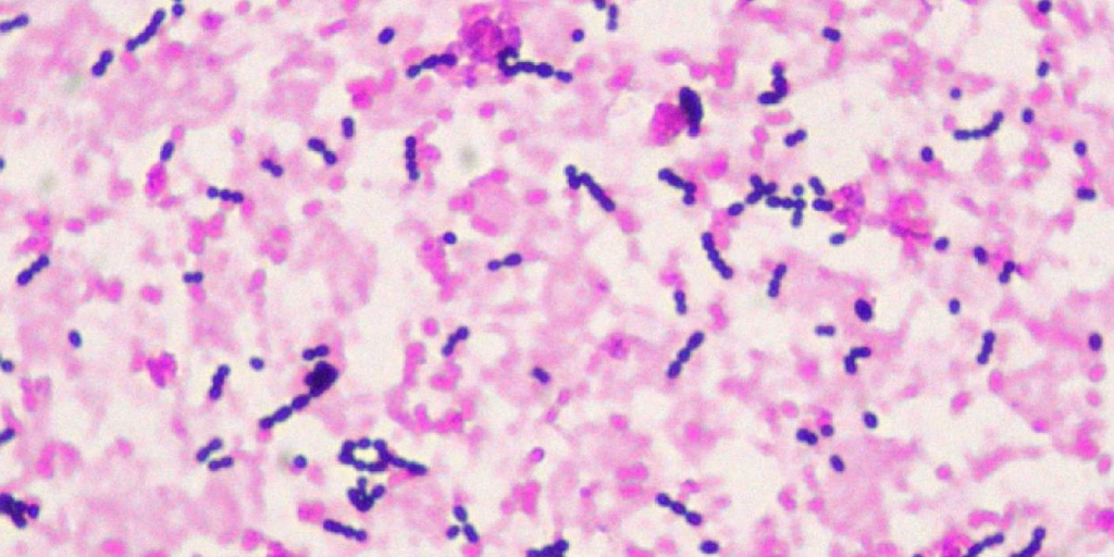
Gram staining is used to differentiate bacteria based on cell wall properties. It differentiates gram positive and gram negative bacteria. Gram positive bacteria retains the primary stain (crystal violet) and appears purple under the microscope while gram negative bacteria retain the secondary stain (Safranin) and appears pink under the microscope.
Reagents Needed
- Crystal violet
- Iodine
- Safranin
- 95% Ethanol.
Procedures
- Prepare a heat fixed smear of the organisms from 24 h old culture on a clean glass slide.
- Flood the smear with crystal violent for 60 seconds
- Rinse the stain off with distilled water
- Flood the smear with Gram’s iodine(mordant) for 30 seconds
- Tilt the slide and rinse with 70% ethanol (decolourizer)
- Flood the smear with Safranin for 60 seconds before rinsing with water
- Blot the slide dry or allow to air dry
- Examine the slide under the microscope with X100 objective lens after adding a drop of immersion oil
References
- Keiser, G.E. ‘Laboratory manual’
- Cain, D., Hanks, H., Weis, M., Bottoms, C., and Lawson J. ‘Microbiology Laboratory Manual Biol 2421L Revised Spring Edition’
Download “Gram Stain”
Gram-Stain.docx – Downloaded 1 time – 20.90 KB


