Introduction
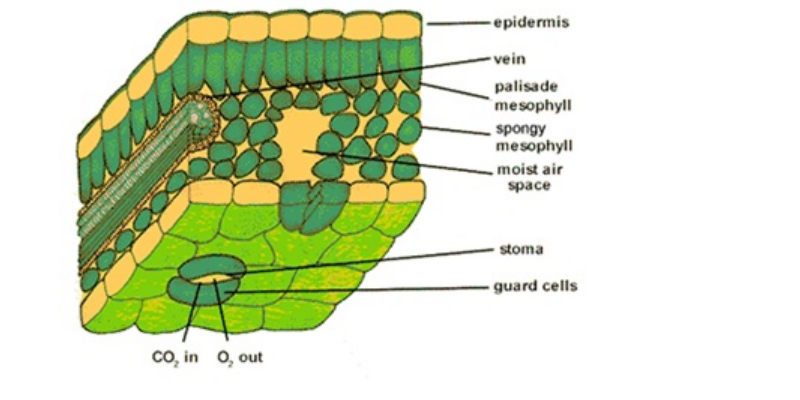
Leaf anatomy may influence net leaf photosynthesis to a large degree and thus cause great differences in light-use efficiency. Like the root and stem, the leaf consists of vascular, parenchymatous and dermal tissue, the latter being a persistent epidermis. The epidermis protects the leaf against uncontrolled high water loss and against damage caused by environmental factors such as wind or micro-organisms. Even though the leaf has a compact arrangement of cells yet there is exchange of gases with the atmosphere through the stomata pores bounded by guard cells which contain chloroplast cells. The vascular bundles, called veins are embedded in the mesophyll and usually surrounded by bundle sheath which may not contain chloroplast cells.
Within the leaf, there is a layer of cells called the mesophyll. Mesophyll can then be divided into two layers, the palisade layer (D) and the spongy layer (F). Palisade cells are more column-like, and lie just under the epidermis, the spongy cells are more loosely packed and lie between the palisade layer and the lower epidermis. The air spaces between the spongy cells allow for gas exchange. Mesophyll cells (both palisade and spongy) are packed with chloroplasts, and this is where photosynthesis actually occurs.
Specimens
The leaf structure of C3 and C4 plants shows clear anatomical and ultrastructural differences: in the C3 pathway plants, the bundle sheath cells are without chloroplast cells whereas chloroplast cells are found in bundle sheath cell of the C4 pathway plants.
The bundle sheath cells which are well supplied with chloroplast cells that often show a reduced granal size synthesise large amounts of starch during the normal photoperiod.
The bundle sheath chlorenchyma is surrounded by a radially arranged mesophyll cell being in direct contact with the bundle sheath. The concentric arrangement of chlorenchyma termed the “kranz-syndrome” or “crown formation” is essential for the functioning of the C4 pathway and is thus characteristic of almost all C4 plants. In the C4 pathway plants, phospho-enol pyruvate carboxylase fixes carbon dioxide in the cytoplasm of the mesophyll cells to form oxalocatate which is further reduced to malate or transaminated to aspartate. These substances are transported into the bundle sheath cells where CO2 is generated by a decarboxylation process and refixed by ribulose 1, 5-diphosphate carboxylase. The initial fixation by PEP carboxylase in the mesophyll cells is a mechanism for concentrating CO2 in the bundle sheath cells to inhibit RUDP oxygenation. This allows RUDP carboxylase to work efficiently even with low CO2 concentration in the intercellular spaces of the mesophyll.
This combination of anatomical and metabolic features suits C4 plants to hot and arid habitats with high irradiation; it may also be an adaptation to short vegetation periods, but only when in conjunction with optimal growing conditions.
Like any other multicellular living thing, a leaf is made up of layers of cells. Viewing the leaf structure under the microscope shows different types of cells that serve various functions. Using a microscope, it’s possible to view and identify these cells and how they are arranged (epidermal cells, spongy cells etc). A diagram of a typical leaf is shown below. It is cut in both longitudinal and cross section to show the cellular structure within.

Terms to be familiar with in the Structure
- Cuticle: This is the outer thick waxy covering of the plants and leaves. Cuticle protects plant from drying out by reducing water loss. It does not let oxygen or carbon dioxide pass through it.
- Upper epidermis: The upper epidermis consists of a single layer of cells covered by cuticle. Sometime contains pores which allow gas or water molecule to pass.
- Veins: It support the lamina and transport materials to and from the leaf tissues, radiate through the lamina from the petiole.
- Stomata: this is an opening between guard cells for gas & water exchange.
- Spongy Mesophyll: This is the lower layer of chloroplast containing cells. They have air spaces around them.
- Palisade Mesophyll: This is the tightly packed upper layer of chloroplast containing cells.
- Guard Cells: 2 cells surrounding stomata that control rate of gas & water exchange.
Activities
View the internal leaf structure of a C3 and C4 plant under the microscope. Make a large labeled diagram of both. Compare C4 leaf structure to that of C3.
References
- ‘Leaf Anatomy in Relation to C3 and C4 Pathways of CO2 Fixation’. Obafemi Awolowo University Botany Laboratory Manual.
- ‘Part 3. Structure of the Plant Leaf Cnd Chloroplasts’ http://projects.ncsu.edu/project/bio183de/Lab/photosynthesis_lab/photosynthesis2H.php Assessed on July 21, 2018.
- ‘Leave Coloring’ https://www.biologycorner.com/worksheets/leaf_coloring.php
- Leaf Structure Under the Microscope Preparation, Requirements and Observations Assessed on July 21, 2018.
- http://science.halleyhosting.com/sci/ibbio/plants/notes/intleaf.php Assessed on July 21, 2018.
- Internal Structure of a Leaf https://www.slideshare.net/LEONARD_AKO/internal-structure-of-a-leaf Assessed on July 21, 2018.
Download “Leaf Anatomy in Relation to C3 and C4 Pathways of CO2 Fixation”
Leaf-Anatomy-in-Relation-to-C3.docx – Downloaded 0 times – 60.59 KB